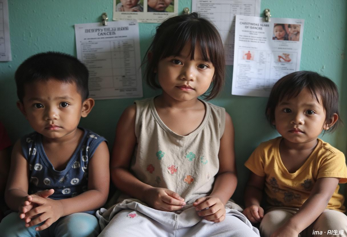
Yolk Sac Tumor
Yolk Sac Tumor
Summarize
1. General remarks
● Overview: Yolk sac tumor is a highly malignant germ cell tumor, which is a kind of non-germ cell germ cell tumor with the differentiation characteristics of extraembryonic yolk sac.
● Manifestation: the specific symptoms are related to the location, size and growth rate of the tumor.
● Treatment: According to the location and stage of the tumor, a combination of surgery, radiotherapy and chemotherapy should be used.
● Prognosis: If diagnosed early and treated properly, the survival rate can reach more than 90%. However, if not treated in time, it may progress rapidly, easy to recur and metastasize, and the survival rate is low.
2. Disease definition
The yolk sac tumor, also known as endodermal sinus tumor (ESET) or infantile embryonal carcinoma (IEC), is a highly malignant germ cell tumor (GCT) that originates from primitive germ cells. It is a type of non-germ cell germ cell tumor (NGGCT) characterized by differentiation features similar to those of the extraembryonic yolk sac. The tumor is named for its structure, which resembles that of the ovarian cystic endodermal sinus in rats and originates from the primitive yolk sac.
Echimycosis may occur in the gonads (testis, ovary) or outside the gonads. Echimycosis outside the gonads is relatively rare and often occurs in the midline of the body. In infants and young children, it is commonly found in the sacrococcygeal region and intracranial, and in adults, it can be seen in the anterior mediastinum, retroperitoneum and intracranial.
3. Epidemiology
Echimycosis can occur at any age, but it is more common in children and adolescents, and can also affect adults. In males, it is most commonly seen before the age of 2; in females, it is most commonly seen before the age of 40, affecting individuals from infancy to adulthood. In children, echimycosis is primarily a simple form, whereas in adults, it often presents as a mixed germ cell tumor that includes other germ cell tumors.
4. Disease type
1) Type of disease
Echimycosis can be classified according to the site of occurrence into intracranial echimycosis, gonadal echimycosis and extra-cranial extra-gonadal echimycosis. Gonadal echimycosis can be further classified into testicular echimycosis and ovarian echimycosis.
2) Disease staging
I) Staging of intracranial yolk sac tumor
Intracranial yolk sac tumors in children can be staged according to the modified Chang staging system for germ cell tumors of the central nervous system in children:
● Limited phase (M0 phase)
A single intracranial lesion with negative cerebrospinal fluid tumor cells; or a double lesion tumor with negative cerebrospinal fluid tumor cells.
● Transfer phase (M+ phase)
The following conditions are considered as the transfer period:
● Non-dual lesion tumors, but more than one intracranial lesion.
● Tumor metastasis to the spinal cord.
● Tumors have metastasized outside the central nervous system.
● Positive for cerebrospinal fluid tumor cells.
ii) Staging of ovarian yolk sac tumor
Pediatric ovarian yolk sac tumors can be staged according to the American Collaborative Group on Childhood Oncology (COG) staging system for pediatric ovarian germ cell tumors:
● Stage I: The tumor is confined to one or both ovaries; complete resection with no ulceration, and negative margins under microscopic examination; the peritoneum is negative, and the peritoneal lavage fluid contains no malignant tumor cells; clinical, radiological, or histological examinations do not reveal any lesions outside the ovaries; tumor markers decrease rapidly to normal levels after surgery due to their half-life decay; the presence of peritoneal neuroglioma does not affect the tumor staging.
Stage ● II: The tumor is confined to one or both ovaries; complete resection without ulceration, with positive margins under microscopic examination; the peritoneal lavage fluid contains no malignant tumor cells, and the peritoneal biopsy is negative; clinical, radiological, or histological examinations do not reveal any lesions outside the ovaries; postoperative serum tumor markers do not show a decrease in their half-life or are abnormal; the presence of peritoneal neuroglioma does not elevate the tumor stage.
● Stage III: gross residual lesions or biopsy only; lymph node involvement, metastatic nodules; visceral involvement (reticulum, intestine, bladder); positive peritoneal biopsy; malignant tumor cells detected in ascites or peritoneal lavage fluid; positive or negative tumor markers.
● Stage IV: distant metastasis, including liver, lung, brain, bone.
iii) Staging of testicular yolk sac tumor
American Collaborative Group on Childhood Tumors (COG) staging system for pediatric testicular germ cell tumors:
● Stage I: The tumor is confined to the testis. A high inguinal incision is made, and the spermatic cord is ligated at the internal ring of the inguinal canal without rupture. The testicular resection is then performed in a direction toward the scrotum. Clinical, radiological, or histological examinations show no lesions outside the testis. Postoperative tumor markers decline rapidly to normal levels due to their half-life. If the patient's tumor markers are normal or undetermined at diagnosis, the lymph nodes in the same side must be negative (if imaging suggests that the lymph node diameter is greater than 2 cm).
● Stage II: Transferred to orifices of the scrotum and testis resection with gross tumor ulceration via a high inguinal incision, followed by peritoneal tumor puncture; microscopic lesions in the scrotum or high spermatic cord (≤5 cm from the proximal end); postoperative serum tumor markers not decreasing or abnormal.
● Stage III: gross tumor remnant; retroperitoneal lymph node metastasis (CT suggests lymph nodes> 4 cm or positive biopsy of lymph nodes> 2cm and <4 cm); visceral or extraperitoneal metastasis.
● Stage IV: distant metastasis, including liver, brain, bone, and lung.
iv) Staging of extra-cranial extra-gonadal yolk sac tumor
Extracranial extra-gonadal yolk sac tumors in children can be staged according to the American Collaborative Group on Childhood Tumors (COG) staging system for extracranial extra-gonadal germ cell tumors in children:
● I stage: complete resection (the sacrococcygeal lesion must be removed from the coccyx), negative margins under the microscope and intact capsule; if the lesion is in the abdominal cavity or retroperitoneum, the tumor cells in ascites are negative; local lymph nodes are histologically negative or imaging suggests <1cm.
● Stage II: total resection with microscopic residual, or biopsy before resection, or capsule rupture; histological negative regional lymph nodes or imaging suggesting <1cm, and tumor cells negative for ascites.
● Stage III: gross residual tumor after resection or only biopsy; histological positivity of regional lymph nodes, or imaging suggesting lymph nodes larger than 2 cm (between 1-2 cm, observation for 4-6 weeks without reduction), and positive tumor cells in ascites.
● Stage IV: distant metastasis including liver, brain, bone and lung.
3) Disease grouping
I) Intracranial yolk sac tumor grouping
The Japanese Pediatric Brain Tumor Study Group (Japanese Pediatric Brain Tumor Study Group) classified intracranial germ cell tumors into good prognosis group, moderate prognosis group and poor prognosis group according to prognosis, and intracranial yolk sac tumor was classified as poor prognosis group.
ii) Extracranial yolk sac tumor grouping
Extracranial yolk sac tumors in children can be classified according to the risk group of extracranial malignant germ cell tumors in children by the Children's Cancer Group (COG) as follows:
Low-risk group: stage I testicular tumor
Medium-risk group: stage II-IV testicular tumors, stage I-III ovarian tumors, and stage I-II extracranial extra-gonadal tumors
High-risk group: stage IV ovarian tumor, stage III-IV extracranial and extra-gonadal tumor
Epidemiological
not have
Etiology & Risk Factors
1. General remarks
The cause of yolk sac tumor is not clear.
2. Basic etiology
Ectodermal sinus tumors originate from primitive germ cells, though the exact cause remains unclear. The prevailing theory suggests that during human development, these germ cells need to migrate to the gonads (ovaries and testes) for further development. However, some of these germ cells make a mistake and fail to migrate properly, instead remaining in the cranium in a relatively primitive state, capable of continuous division and proliferation, eventually leading to the formation of germ cell tumors. However, this theory still requires further scientific evidence to support it.
3. Triggering factors
Studies have shown that children with undescended testicles are more likely to develop testicular germ cell tumors (including yolk sac tumors) than the general population.
Classification & Staging
not have
Clinical Manifestations
1. General remarks
The symptoms of the patient are related to the location, size and growth rate of the yolk sac tumor.
2. Typical symptoms
Depending on where the tumor grows, a patient's symptoms can vary widely.
The symptoms of intracranial yolk sac tumor depend on the tumor's location. Common symptoms include increased intracranial pressure, which can cause headaches, vomiting, papilledema, drowsiness, ataxia, and behavioral changes; hypothalamic/pituitary dysfunction, such as diabetes insipidus, delayed puberty or precocious puberty, simple growth hormone deficiency, central hypothyroidism, and adrenal insufficiency; and ocular abnormalities, such as difficulty in upward gaze, convergence nystagmus, decreased vision, and visual field defects.
Patients with ovarian yolk sac tumors often present with abdominal pain and pelvic masses. They may be accompanied by severe pain, which can easily be misdiagnosed as appendicitis. The tumor may grow very rapidly and aggressively, spreading widely in the peritoneum.
Testicular yolk sac tumors typically present as nodules or painless swelling in one testicle. Some patients may experience dull pain or a feeling of heaviness in the lower abdomen, perianal area, or scrotum, with a few experiencing acute pain. In a small number of cases, the initial symptoms of testicular tumors are due to metastatic disease, which can vary depending on the site of metastasis, including neck masses (metastasis to supraclavicular lymph nodes), coughing or breathing difficulties (lung metastasis), loss of appetite, nausea, vomiting, or gastrointestinal bleeding (post-duodenal metastasis), back pain (massive retroperitoneal space-occupying lesions involving lumbar muscles or nerve roots), and bone pain (bone metastasis).
Thyrogastromegaly grows rapidly and may cause fever, chills, weight loss, chest pain, dyspnea, and/or superior vena cava syndrome.
The main manifestation of sacrococcygeal yolk sac tumor is a mass at the end of the patient, which may be asymptomatic or accompanied by rectal or bladder obstruction, pain and other symptoms.
Clinical Department
not have
Examination & Diagnosis
1. General remarks
The gold standard for diagnosing yolk sac tumors is histopathological diagnosis. If a biopsy is not available, clinical diagnostic criteria can be used based on imaging and tumor marker levels.
2. Department of treatment
Intracranial yolk sac tumors can be treated in pediatric neurology or pediatric neurosurgery, as well as neurology or neurosurgery.
Extracranial yolk sac tumor can be treated in pediatric oncology, pediatric oncology surgery, oncology, oncology surgery and other departments.
3. Diagnostic basis
Pathological diagnosis is the gold standard for diagnosing yolk sac tumor. Pathological diagnosis is usually made by surgical resection or excision of tumor tissue, or by obtaining tumor tissue samples by biopsy.
If the risk of surgical resection or biopsy of tumor tissue is high, clinical diagnosis can be adopted with the following criteria:
● There are clear imaging characteristics of yolk sac tumor.
● Alpha-fetoprotein (AFP) levels are above normal.
It should be noted that because testicular biopsy may lead to tumor dissemination into the scrotum or metastasis to the inguinal lymph nodes, testicular yolk sac tumors are usually not diagnosed by biopsy but by clinical diagnostic criteria.
4. Related checks
1) Physical examination
General physical signs are checked for signs of disease, such as lumps or any other unusual manifestations. The patient's health habits, past illnesses and treatment history are also recorded.
The physical examination includes abdominal palpation to see if there is evidence of nodular lesions or visceral involvement; the doctor also usually evaluates supraclavicular lymph nodes to detect patients with advanced disease with enlarged lymph nodes. The chest examination can detect whether the patient may have chest involvement.
For patients suspected of having testicular tumors, a palpation examination of the testicles is required.
2) Imaging examination
I) Chest X-ray (chest radiograph) examination
Chest radiography can be used as an initial imaging test for mediastinal tumors, lymph node metastasis and lung metastasis.
ii) CT plain scan examination
CT plain scan is sensitive to intracranial tumors, especially those in the pineal gland and suprasellar regions where yolk sac tumors often occur. It can determine the location and size of lesions, clarify calcification, cystic changes or bleeding and the degree of hydrocephalus, and judge the affected area, but cannot determine the type of tumor.
High resolution CT of abdomen and pelvis is helpful in detecting gonadal or mediastinal tumors and lymph node metastasis.
Chest CT is recommended if the chest radiograph results are abnormal or there is a high suspicion of metastatic lesions involving the chest.
iii) Magnetic Resonance Imaging (MRI)
Magnetic resonance imaging is more sensitive than CT in tumor diagnosis and staging, can more clearly show the location and range of lesions, the relationship between lesions and adjacent structures, the anatomical structure of adjacent vascular system, and can better find distant disseminated lesions. However, magnetic resonance imaging is less sensitive than CT in showing calcification.
Iv) B-ultrasound examination
For gonadal tumors, B-ultrasound can help determine the exact location of the lesion.
3) Tumor marker examination
Germ cell tumors often secrete tumor markers such as alpha-fetoprotein (AFP), lactate dehydrogenase (LDH), and human chorionic gonadotropin β-subunit (β-hCG). Testing these markers in the blood can help differentiate between different types of germ cell tumors. These serum markers need to be tested through a blood draw.
Ectodermal cystomas can secrete alpha-fetoprotein. In children, the normal level of alpha-fetoprotein is usually 0-25μg/L (which varies by age).
4) Histopathological examination (biopsy or surgical resection specimen)
Tumor samples for histopathological examination can be obtained through tissue biopsies or surgical resections. For intracranial tumors, a biopsy involves removing a portion of the skull and using a fine needle to extract tissue samples for examination, to identify and determine the type of tumor cells. For tumors in other areas, histopathological examination can be performed through puncture, surgical resection, or excision of tissue samples.
The small sample size of a biopsy can sometimes lead to an inaccurate diagnosis. The larger sample size of a surgical resection is more conducive to an accurate diagnosis, but it is also more invasive to the body.
The results of histopathological examination are the most important diagnostic basis for the diagnosis of yolk sac tumor. However, testicular biopsy may lead to the spread of the tumor into the scrotum or metastasis and spread to the inguinal lymph nodes, so testicular yolk sac tumor is usually not diagnosed by biopsy, but by clinical diagnostic criteria.
5) Cerebrospinal fluid cytology examination (lumbar puncture examination)
For intracranial tumors, if the patient is suitable for a lumbar puncture (LP), it is advisable to perform a cerebrospinal fluid cytology examination to stage the tumor. The cerebrospinal fluid cytology examination requires a sample taken through the lumbar puncture. Pathologists will examine the cerebrospinal fluid sample under a microscope for signs of tumor cells, which can help stage germ cell tumors.
At the same time, for intracranial tumors, the tumor marker alpha-fetoprotein in cerebrospinal fluid is often more sensitive than serum level, so the level of tumor markers in cerebrospinal fluid can be checked at the same time to assist diagnosis.
5. Differential diagnosis
The differential diagnosis of testicular yolk sac tumor includes testicular torsion, epididymitis or orchitis, and some less common conditions such as hydrocele, varicocele, hernia, hematoma, and spermatocele. For patients with unclear diagnoses or those whose hydrocele hinders a thorough examination, imaging tests can help determine the cause.
Intracranial yolk sac tumors require careful differentiation from other brain tumors in the pineal region and suprasellar area, such as pineal germ cell tumors, ependymomas of the pineal region, craniopharyngiomas, Langhans cell histiocytosis of the suprasellar region, low-grade gliomas, hamartomas, or brain metastases from extracranial tumors.
Clinical Management
1. General remarks
Echocystoma usually requires a combination of multiple treatments. Intracranial echocystoma usually requires surgery combined with radiotherapy and chemotherapy, while extracranial echocystoma requires surgery combined with chemotherapy.
2. General treatment
Intracranial yolk sac tumors are typically treated with a combination of multiple drug chemotherapy, whole brain and spinal cord radiotherapy, and tumor bed radiotherapy. Residual lesions after radiotherapy are surgically removed. The specific radiotherapy dose and surgical plan are determined based on the effectiveness of the chemotherapy. For patients under 3 years old, to avoid the side effects of radiotherapy on the central nervous system's development, radiotherapy is delayed until the patient is 3 years old, with chemotherapy and surgery being performed first.
Testicular stage I yolk sac tumor can be cured by orchiectomy alone, and follow-up observation is required without further treatment. Stage II to IV requires surgery and postoperative chemotherapy; if the surgical evaluation is difficult to completely remove, neoadjuvant chemotherapy should be performed before surgery.
Ovarian yolk sac tumors require surgery and postoperative chemotherapy; if the surgical evaluation is difficult to completely remove, neoadjuvant chemotherapy should be performed before surgery.
The main treatment for extracranial extra-gonadal yolk sac tumor is surgery and postoperative chemotherapy. For extracranial extra-gonadal yolk sac tumor in stage III-IV, if the surgical evaluation is difficult to completely resect, neoadjuvant chemotherapy should be performed before surgery.
3. Chemotherapy
1) Chemotherapy regimen
I) Chemotherapy regimen for intracranial yolk sac tumor
For intracranial yolk sac tumors, preoperative chemotherapy is required. Typically, this involves alternating between the EP regimen (epirubicin + etoposide), the CE regimen (carboplatin + etoposide), and the IE regimen (ifosfamide + etoposide) for at least six cycles. Before chemotherapy, the patient must have an absolute neutrophil count (ANC) above 1000/μl, platelet count above 100,000/μl, and normal liver and kidney function. After chemotherapy, granulocyte colony-stimulating factor (G-CSF, commonly known as 'white blood cell booster') should be administered.
ii) Chemotherapy regimen for extra-cranial yolk sac tumor
The preferred chemotherapy regimen for extracranial yolk sac tumors is the BEP regimen (Bortezomib + Etoposide + Paclitaxel). Considering the ototoxic and nephrotoxic effects of Paclitaxel, the JEB regimen (Bortezomib + Etoposide + Carboplatin) can also be considered. However, studies have shown that the survival rate with the JEB regimen for testicular germ cell tumors is lower than with the BEP regimen, so it is advisable not to easily replace Paclitaxel with Carboplatin in the early stages of treatment. If the BEP or JEB regimens are ineffective, a 4-drug regimen can be added to the BEP or JEB regimens, including doxorubicin, ifosfamide, cyclophosphamide, vincristine, or actinomycin D. Alternatively, a combination regimen of cyclophosphamide, Paclitaxel, vincristine, actinomycin D, and bleomycin, or vincristine, bleomycin, Paclitaxel, cyclophosphamide, actinomycin D, and doxorubicin can be tried. For refractory extracranial yolk sac tumors, a combination regimen of paclitaxel, ifosfamide, and Paclitaxel/Carboplatin can be considered; or an irinotecan monotherapy or irinotecan in combination with Paclitaxel/Nadaplatin can be attempted.
The low-risk group typically requires 3-4 chemotherapy courses after tumor resection or once serum tumor marker levels return to normal post-surgery. The intermediate-risk group needs 6 chemotherapy courses after tumor resection or 4 courses once serum tumor marker levels return to normal post-surgery. The high-risk group requires 8 chemotherapy courses after tumor resection or 4 courses once serum tumor marker levels return to normal post-surgery.
2) Adverse reactions
Common adverse reactions include bone marrow suppression (manifested as neutropenia, thrombocytopenia, anemia, etc.), nephrotoxicity (cisplatin), nausea and vomiting, allergic reaction, hair loss, hearing damage caused by ototoxicity (cisplatin), fatigue, electrolyte disturbance, neurotoxicity, etc.
4. Radiotherapy
1) Radiotherapy regimen
Egnetoma often has a poor response to radiotherapy. Some intracranial egnetomas may require whole brain and spinal cord radiotherapy and tumor bed radiotherapy, while extracranial egnetomas are generally not treated with radiotherapy.
2) Adverse reactions
The side effects of radiotherapy include nausea and vomiting. Radiotherapy to brain tissue may affect the neurological development of children. Whole-brain and whole-spinal cord radiotherapy can increase the incidence of neurological complications, including neurocognitive impairments. For developing children, it may also lead to delayed bone growth, hypothyroidism, adrenal insufficiency, and hypogonadism, requiring long-term follow-up by pediatric health care and endocrinology departments to mitigate the side effects of treatment.
5. Surgical treatment
Egret sac tumor needs to be removed by surgery, and the surgery can also take samples to confirm the histopathological diagnosis, so as to determine the stage of the disease and subsequent treatment plan.
For ovarian tumors, ovarian resection, ovarian cystectomy or ovarian mass resection may be performed according to the condition. For testicular tumors, radical inguinal orchiectomy and retroperitoneal lymph node dissection are required.
For infantile vaginal yolk sac tumor, conservative surgery is usually the first choice. The first operation is to remove the tumor and perform biopsy. In the second operation after chemotherapy, vaginal preservation surgery is performed and residual disease is removed as thoroughly as possible.
Prognosis
1. General remarks
If the child's yolk sac tumor can be diagnosed early and treated in a standardized way, according to the statistics of the United States, the survival rate can reach more than 90%. However, due to the high malignancy of yolk sac tumor, if not treated in time, it may progress rapidly, easy to recur and metastasize, and the survival rate is low.
There are many factors affecting the prognosis, including clinical stage, response to chemotherapy drugs, and standardization of treatment and follow-up.
2. Aftereffects
For tumors in the gonadal area, one side of the gonad may need to be removed, which could affect the child's sexual development. Currently, there is no cure. Regular monitoring of the healthy ovary or testicle is necessary. For younger children, it is important to check the levels of sex hormones (estrogen and testosterone) after they reach puberty. If these hormone levels are insufficient, they can be supplemented externally to ensure the child's normal development.
Platinum-based chemotherapy is ototoxic in children and can cause irreversible hearing loss if taken in excess.
In addition, if the patient receives whole brain and spinal cord radiotherapy, the incidence of neurological complications (including neurocognitive dysfunction) will increase. Irradiation to the nervous system may cause neurocognitive dysfunction, which will affect attention, memory and processing ability, etc., depending on the age and dose of radiotherapy at the time of radiotherapy.
Radiation therapy may also increase the risk of developing a second tumor.
3. transition
Germ cell tumors are more likely to involve lymph nodes, with lymph node metastasis occurring in approximately 28% of cases (including children and adults).
Follow-up & Review
not have
Daily Care
1. General remarks
After the completion of yolk sac tumor treatment, it is important to carry out timely review and follow-up, on the one hand to monitor the side effects caused by treatment, on the other hand to pay close attention to whether the disease is recurrent.
2. Reexamination
Follow-up should be paid attention to at least 5 years after the end of treatment, mainly tumor marker examination and imaging examination. Lifelong follow-up is recommended.
Within 3 months, the level of tumor marker alpha-fetoprotein was rechecked every half month.
Tumor marker alpha-fetoprotein levels were rechecked once a month for 3 months to less than 1 year.
One year later, the level of the tumor marker alpha-fetoprotein was rechecked every two to three months.
Imaging [B-ultrasound and/or CT or magnetic resonance imaging (MRI)] is performed every 3 months to examine the primary site and sites of metastasis (e.g., liver, lung, and draining lymph nodes).
3. Daily life management
1) Diet
It is important to provide patients with a nutrient-rich and balanced diet, ensuring the intake of high-quality proteins such as meat, eggs, dairy, poultry, fish, shrimp, soybeans, and soy products. Additionally, patients should consume more whole grains, vegetables, and fruits, and moderately eat dairy products and nuts to ensure the intake of other essential nutrients. Patients can consult clinical nutritionists from the hospital's nutrition department for a suitable nutritional plan. If there is a significant weight loss, consider using tube feeding or parenteral nutrition for nutritional support.
2) Movement
It is important to ensure that patients get enough sleep. Regular and quality sleep is very helpful for physical recovery and immunity. A suitable sleep environment (usually dark, quiet and at a comfortable temperature) may help improve the quality of sleep.
If the patient's physical condition allows, encourage and assist the patient to do some simple activities. Moderate exercise is helpful in preventing muscle atrophy, enhancing physical strength and endurance, and promoting appetite.
3) Lifestyle
If the patient is caused by treatment of neutropenia, attention should be paid to prevent infection. Pay attention to personal and living environment hygiene, do not approach patients with infectious diseases, and do not go to crowded places.
If the treatment causes thrombocytopenia, you should avoid bleeding. You should stay away from sharp, spiky toys and objects, and avoid strenuous sports (such as jumping, soccer, basketball, etc.).
4. Daily disease monitoring
Postoperative complications, chemotherapy side effects (such as hair loss, fatigue, vomiting, etc.), tumor metastasis and recurrence, growth and development problems should be paid attention to. When fever, worsening symptoms, new symptoms and treatment side effects occur, consult your doctor in time.
5. Special Precautions
Children with yolk sac tumors who have undergone radiotherapy and chemotherapy are at risk of long-term side effects and secondary cancers, which may occur many years after the end of treatment, and this risk is related to the regimen and dose used during treatment. Therefore, all medical visits and treatment records should be kept so that they can be used as a reference for future review and medical care.
6. Prevention
Because the exact cause of yolk sac tumor is not clear, there is no good way to prevent the occurrence of yolk sac tumor. However, regular follow-up and maintaining a good healthy lifestyle can help prevent and detect the recurrence or long-term effects of the disease as early as possible.
Cutting-edge therapeutic and clinical Trials
not have
References
1. Chinese Anti-Cancer Association Pediatric Oncology Committee. Expert Consensus on Multidisciplinary Diagnosis and Treatment of Primary Germ Cell Tumors of the Central Nervous System in Children. Chinese Journal of Pediatric Hematology and Oncology. 2018.23(6):281-286.
2.Su, JM. Intracranial germ cell tumors. In: UpToDate, Eichler, AF, Loeffler, JS, Wen, PY, Gajjar, A, (Ed), UpToDate, Waltham, MA, 2020.
3.PDQ® Pediatric Treatment Editorial Board. PDQ Childhood Central Nervous System Germ Cell Tumors Treatment (Health Professional Version). Bethesda, MD: National Cancer Institute. Updated <12/17/2019>. Available at: https://www.cancer.gov/types/brain/hp/child-cns-germ-cell-treatment-pdq. Accessed <06/08/2020>. [PMID: 26389498]
4.PDQ® Pediatric Treatment Editorial Board. PDQ Childhood Central Nervous System Germ Cell Tumors Treatment (Patient Version). Bethesda, MD: National Cancer Institute. Updated <02/05/2020>. Available at: https://www.cancer.gov/types/brain/patient/child-cns-germ-cell-treatment-pdq. Accessed <06/08/2020>. [PMID: 26389502]
5.PDQ® Pediatric Treatment Editorial Board. PDQ Childhood Extracranial Germ Cell Tumors Treatment. Bethesda, MD: National Cancer Institute. Updated <05/28/2020>. Available at: https://www.cancer.gov/types/extracranial-germ-cell/hp/germ-cell-treatment-pdq. Accessed <06/19/2020>. [PMID: 26389316]
6.PDQ® Pediatric Treatment Editorial Board. PDQ Childhood Extracranial Germ Cell Tumors Treatment. Bethesda, MD: National Cancer Institute. Updated <02/10/2020>. Available at: https://www.cancer.gov/types/extracranial-germ-cell/patient/germ-cell-treatment-pdq. Accessed <06/19/2020>. [PMID: 26389180]
7. Expert consensus on multidisciplinary diagnosis and treatment of extracranial germ cell tumors in children (CCCG-GCTs-2018).2019.
8.Steele, GS, Richie, JP, Oh, WK, Michaelson, MD. Clinical manifestations, diagnosis, and staging of testicular germ cell tumors. In: UpToDate, Kantoff, PW, Shah, S, (Ed), UpToDate, Waltham, MA, 2020.
9.Gershenson, DM. Ovarian germ cell tumors: Pathology, epidemiology, clinical manifestations, and diagnosis. In: UpToDate, Goff, B, Pappo, AS, Garcia, RL, Chakrabarti, A,(Ed), UpToDate, Waltham, MA, 2020.
10.Kantoff, PW. Extragonadal germ cell tumors involving the mediastinum and retroperitoneum. In: UpToDate, Oh, WK, Shah, S, Hollingsworth, H, (Ed), UpToDate, Waltham, MA, 2020.
11. Bao Nan, Zhang Xiaolun and Liu Jing. Diagnosis and treatment of pediatric yolk sac tumor. Chinese Journal of Pediatric Surgery. 2011; 32:3.
12.Faure Conter C, Xia C, Gershenson D, et al. Ovarian Yolk Sac Tumors; Does Age Matter? Int J Gynecol Cancer. 2018;28(1):77-84. doi:10.1097/IGC.0000000000001149
13.SFrazier, A. L., Hale, J. P., Rodriguez-Galindo, C., Dang, H., Olson, T., Murray, M. J., … Nicholson, J. C. Revised Risk Classification for Pediatric Extracranial Germ Cell Tumors Based on 25 Years of Clinical Trial Data From the United Kingdom and United States. Journal of Clinical Oncology, 2015. 33(2), 195–201. doi:10.1200/jco.2014.58.3369
Audit specialists
Wang Jingfu, director of the Department of Pediatric Oncology, Shandong Cancer Hospital
Baidu Encyclopedia link: https://baike.baidu.com/item/%E7%8E%8B%E6%99%AF%E7%A6%8F/50993639
Search
Related Articles

Relaxation Therapy & Peace Care
Jul 03, 2025

Rare Childhood Tumour
Jul 03, 2025

Inflammatory Myofibroblastoma
Jul 03, 2025

Langerhans Cell Histiocytosis
Jul 03, 2025

Angeioma
Jul 03, 2025